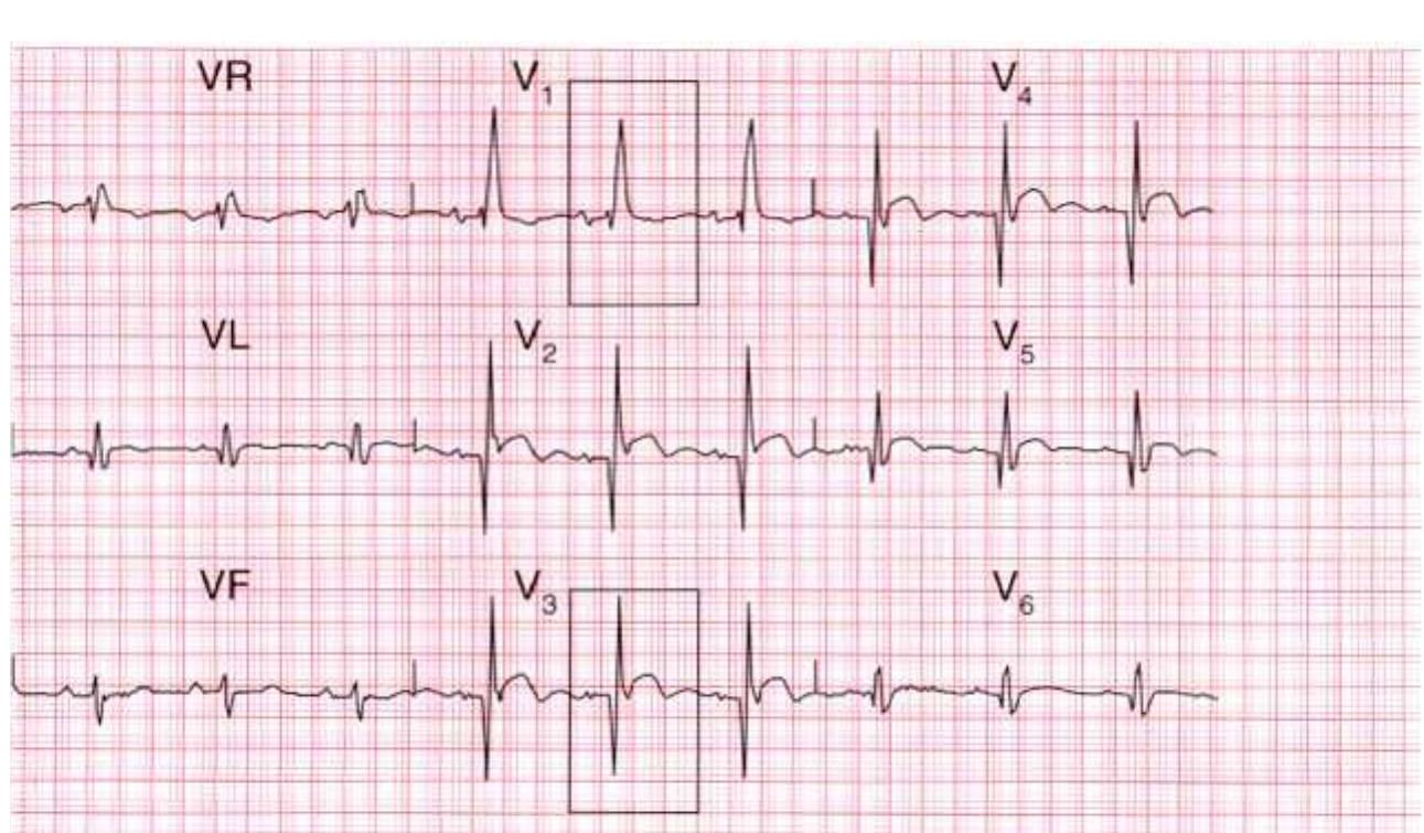
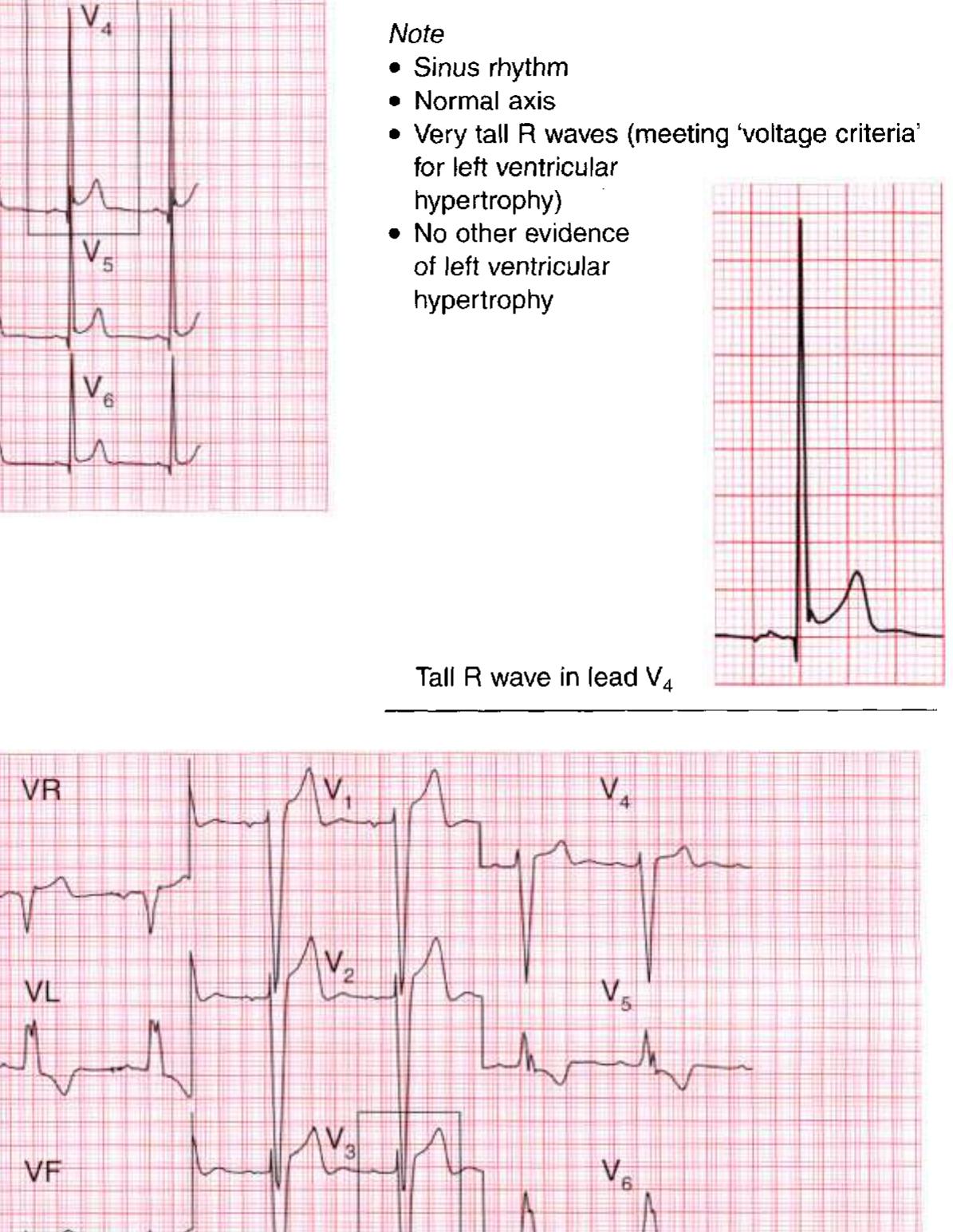
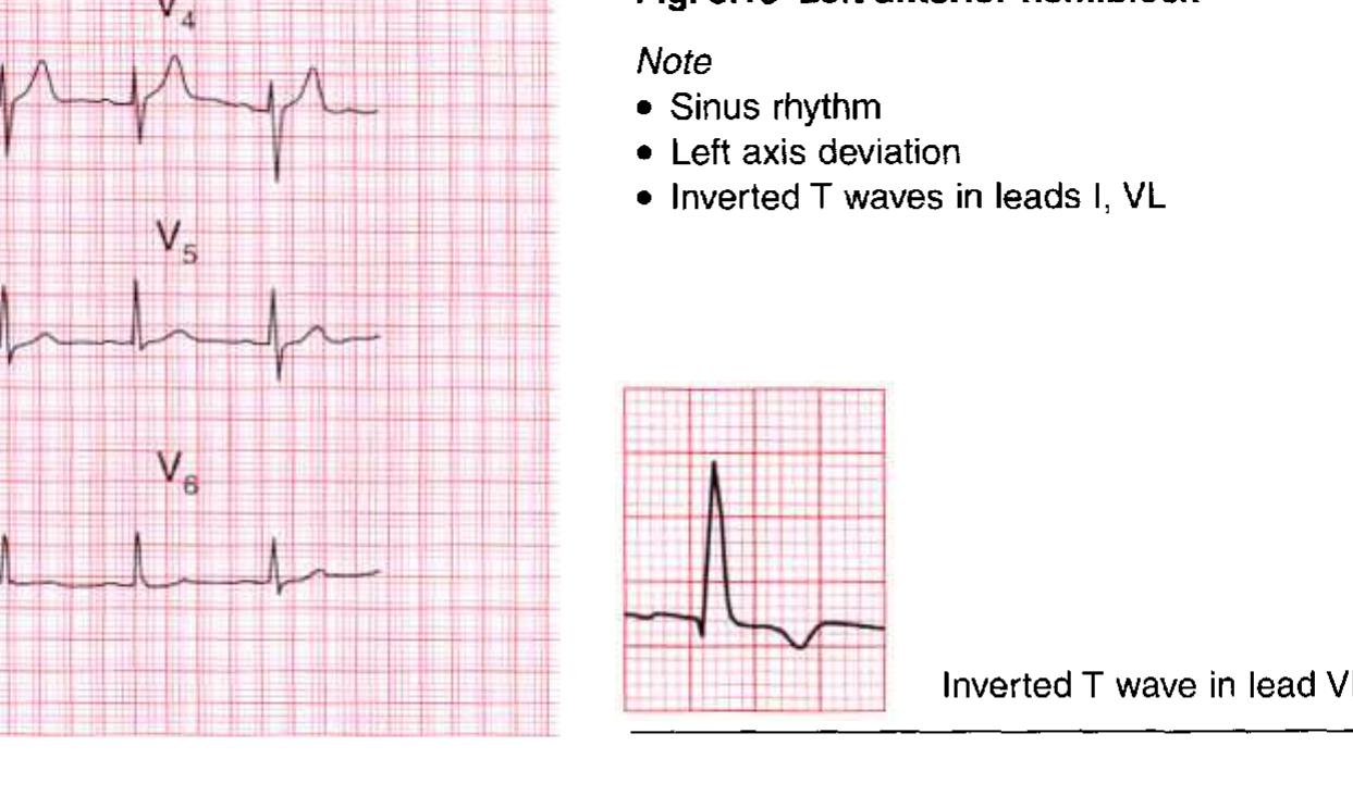
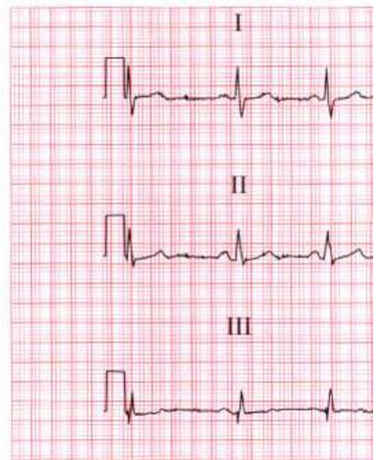
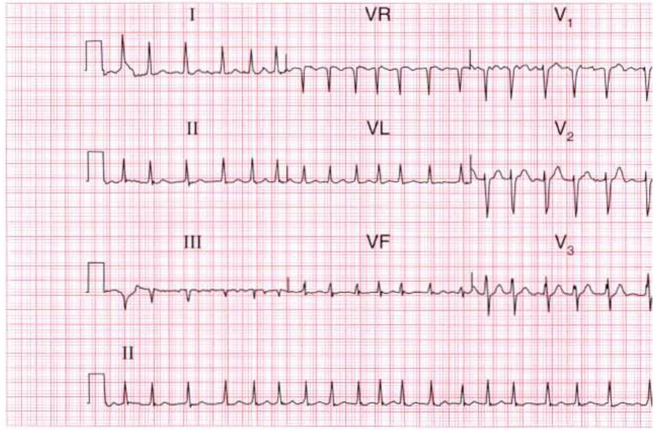
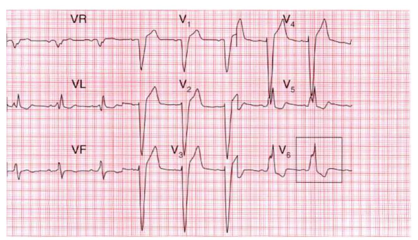
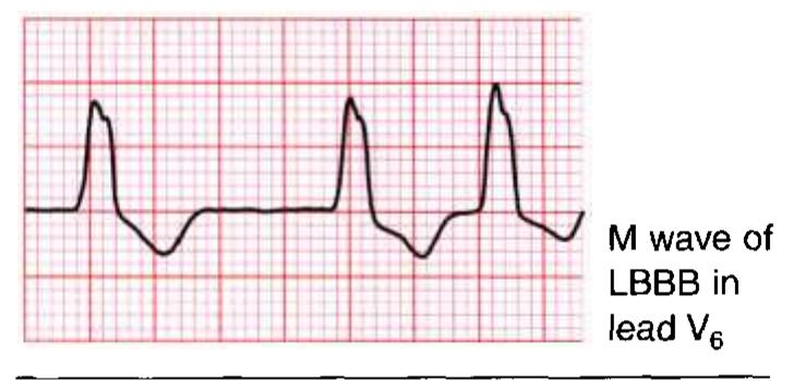
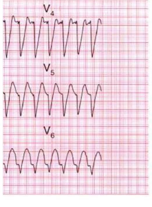
The ECG made easyThis book and the individual contributions contained in it are protected under copyright by the Publisher (other than as may be noted herein). Notices Practitioners and researchers must always rely on their own experience and knowledge in evaluating and using any information, methods, compounds or experiments described herein. Because of rapid advances in the medical sciences, in particular, independent verification of diagnoses and drug dosages should be made. To the fullest extent of the law, no responsibility is assumed by Elsevier, authors, editors or contributors for any injury and/or damage to persons or property as a matter of products liability, negligence or otherwise, or from any use or operation of any methods, products, instructions or ideas contained in the material herein. Part 2: The basics: the fundamentals of ECG recording, reporting and interpretation Before you can use the ECG as an aid to diagnosis or treatment, you have to understand the basics. Part 2 of this book explains why the electrical activity of the heart can be recorded as an ECG, and describes the significance of the 12 ECG 'leads' that make 'pictures' of the electrical activity seen from different directions. Part 2 also explains how the ECG can be used to measure the heart rate, to assess the speed of electrical conduction through different parts of the heart, and to determine the rhythm of the heart. The causes of common 'abnormal' ECG patterns are described. Part 4: Now test yourself You should now be able to recognize the common ECG patterns, and this final chapter contains twelve 12-lead ECGs from real patients for you to interpret. Quick reminders This has been placed at the back of the book after the index so you can refer to it quickly when you need to. It lists the common abnormalities you must be able to recognize. Further reading The symbol indicates cross-references to useful information in The ECG Made Practical, 7th edition (Elsevier, 2019). 'ECG' stands for electrocardiogram, or electrocardiograph. In some How to record an ECG? Electrodes are placed on the chest and limbs of the patient to record different views of the heart's electrical activity. Each view of the heart is described as a 'lead'. The word 'lead' does not refer to the electrodes. The rhythm of the heart can be determined from only one view, i.e. one lead (this requires two electrodes). For a full picture of the heart's electrical activity, a 12-lead view is conventional. One electrode is attached to each limb. These four electrodes provide six 'limb leads' or six different views of the heart in a vertical plane. These are called leads I, II, III, VL, VF and VR. VL, VF and VR used to be called AVL, AVF and AVR, respectively, but the A is essentially meaningless and is redundant. Six electrodes are attached to the chest, recording leads V 1 to V 6. Accurate placement of these electrodes is essential for comparing later ECGs. These leads 'look at' the heart from the front in a horizontal plane (Fig. 1.1). ECG abnormality Consider Ventricular rate above 120 bpm or below 45 bpm Ischaemia, hypotension, sepsis Atrial fibrillation Valve disease, alcoholism, ischaemia, infection Complete heart block Any heart disease ST segment elevation or depression Infarction, ischaemia Abnormal T wave inversion Infarction, ischaemia, pulmonary embolism Wide QRS width Any heart disease ■ TOP TIP: DON'T PANIC-THE ECG REALLY IS VERY EASY! Now you are ready to read the remainder of the book. 'ECG' stands for electrocardiogram, or electrocardiograph. In some countries, the abbreviation used is 'EKG'. Remember: FIG. 2.27 The effect of over-calibration Note Ventricular tachycardia, 74-75, 75f, 76f, 151f, 156f heart rate, diagnosis, 64-67 paroxysmal, 149 pulseless, shockable cardiac arrest, 160 supraventricular tachycardia vs., with bundle branch block, 75-77 torsade de pointes, 149, 151f Voltage changes, effect, 89 Voltage criteria, left ventricular hypertrophy, 89
 ASHRAF ALQUDWA


![S waves in jeads J] and Ill: left axis deviation](https://figures.academia-assets.com/36721212/figure_392.jpg)